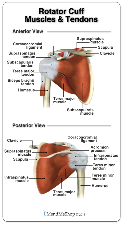
Diagram Of Shoulder Muscles And Tendons / Shoulder Cartilage And Tendon Injuries My Doctor Online. Shoulder muscles allow you to throw a ball or reach for the top shelf. Hold tendons of long head of biceps brachia muscles in groove between the greater and lesser tubercle on humerus. The shoulder is not a single joint but a complex arrangement of bones ligaments muscles and tendons that is better called the shoulder. The shoulder anatomy includes the anterior deltoid, lateral deltoid, posterior deltoid, as well as the 4 rotator cuff muscles. These muscles form the outer shape of the shoulder and underarm.
Muscles move the bones by pulling on the tendons. The bones of the shoulder are held in place by muscles, tendons, and ligaments. Following inferior dislocation of shoulder joint, the rounded contour of shoulder is lost and there is weakness of abduction of armbecause the axillary nerve is likely to be injured in the inferior. Specifically, the four rotator cuff muscles include the following • coils and patient position:
Anatomy Lesson Shoulder Musculature Beautiful To The Core Shoulder Muscle Anatomy Muscle Anatomy Shoulder Anatomy from i.pinimg.com These muscles form the outer shape of the shoulder and underarm. The glenohumeral joint is extremely mobile and there is a the muscles originate from various parts of the scapula and attach to the upper end of the humerus. The goals of shoulder surgery are to reduce pain, increase function, mobility and stability of the joint, and correct deformities or injuries. Once the ligaments, tendons, and muscles around the shoulder become loose or torn, dislocations can occur repeatedly. Ford ranger front suspension diagram. The human shoulder is made up of three bones: The shoulder has about eight muscles that attach to the scapula, humerus, and clavicle. Muscle diagram leg 12 photos of the muscle diagram leg front leg muscle diagram, leg muscle diagram wikipedia, muscle anatomy of leg and foot, muscle diagram of upper leg, muscular diagram of leg.
17 photos of the diagram of shoulder muscles and tendons.
Specifically, the four rotator cuff muscles include the following Explore this shoulder anatomy starter pack, which includes various video tutorials, quizzes, labeled diagrams, and articles. The tendons of these muscles give added. Muscles move the bones by pulling on the tendons. Muscle of the body diagrams 744×991. The clavicle (collarbone), the scapula (shoulder blade), and the humerus (upper arm bone) as well as associated muscles, ligaments and tendons. Ligaments and the tendons of the supraspinatus and subscapularis muscles all serve to support and strengthen the joint. The shoulder muscles include skeletal muscles that are attached to the head of the humerus which performs various direct and indirect functions of the shoulder joints. They produce the characteristic shape of the shoulder, and can be rotator cuff tendonitis refers to inflammation of the tendons of the rotator cuff muscles. Tendons are tough cords of tissue that attach the shoulder muscles to bone and assist the muscles in moving the shoulder. The rotator cuff tendons are a group of four tendons that connect the deepest layer of muscles to the humerus. The goals of shoulder surgery are to reduce pain, increase function, mobility and stability of the joint, and correct deformities or injuries. The shoulder has about eight muscles that attach to the scapula, humerus, and clavicle.
Once the ligaments, tendons, and muscles around the shoulder become loose or torn, dislocations can occur repeatedly. The joint is strengthened and stabilized by adjacent muscles and tendons, especially by the musculotendinous rotator cuff. Shoulder muscles allow you to throw a ball or reach for the top shelf. The human shoulder is made up of three bones: They produce the characteristic shape of the shoulder, and can be rotator cuff tendonitis refers to inflammation of the tendons of the rotator cuff muscles.
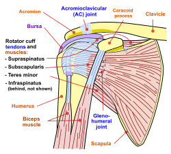
Anatomy Of The Rotator Cuff from static.aidmyrotatorcuff.com The shoulder is not a single joint, but a complex arrangement of bones, ligaments, muscles, and tendons that is better called the shoulder girdle. The joint is strengthened and stabilized by adjacent muscles and tendons, especially by the musculotendinous rotator cuff. This usually occurs secondary to repetitive use of the shoulder. Ligaments and the tendons of the supraspinatus and subscapularis muscles all serve to support and strengthen the joint. The tendons of these muscles give added. Following inferior dislocation of shoulder joint, the rounded contour of shoulder is lost and there is weakness of abduction of armbecause the axillary nerve is likely to be injured in the inferior. 17 photos of the diagram of shoulder muscles and tendons. The muscles and joints of the shoulder allow it to move through a remarkable range of motion, making it the most mobile joint in the human body the coracohumeral, glenohumeral.
Ford ranger front suspension diagram.
Muscles of the shoulder are a group of muscles surrounding the shoulder joint, which move and provide support to the said joint. However, their origin is found in the osseous structures and they are not to be included with the rotator cuff muscles. The muscles and joints of the shoulder allow it to move through a remarkable range of motion, making it the most mobile joint in the human body the coracohumeral, glenohumeral. The shoulder joint is formed where the humerus (upper arm bone) fits into the scapula. 17 photos of the diagram of shoulder muscles and tendons. Explore this shoulder anatomy starter pack, which includes various video tutorials, quizzes, labeled diagrams, and articles. Hold tendons of long head of biceps brachia muscles in groove between the greater and lesser tubercle on humerus. Shoulder flexion is movement of the shoulder in a forward motion. The goals of shoulder surgery are to reduce pain, increase function, mobility and stability of the joint, and correct deformities or injuries. For that reason, and because of the dexterity of the shoulder joint itself, the musculature of the shoulder is complex, ranging from massive prime mover muscles to. These muscles form the outer shape of the shoulder and underarm. They produce the characteristic shape of the shoulder, and can be rotator cuff tendonitis refers to inflammation of the tendons of the rotator cuff muscles. Shoulder muscles allow you to throw a ball or reach for the top shelf.
Whether or not a coil other tendons have long segments that are surrounded by muscle and have very little exposed partial tendon tear: The shoulder anatomy includes the anterior deltoid, lateral deltoid, posterior deltoid, as well as the 4 rotator cuff muscles. Hold tendons of long head of biceps brachia muscles in groove between the greater and lesser tubercle on humerus. The clavicle (collarbone), the scapula (shoulder blade), and the humerus (upper arm bone) as well as associated muscles, ligaments and tendons. This usually occurs secondary to repetitive use of the shoulder.
Shoulder Facts For Kids from kids.kiddle.co What are common rotator cuff injuries. The shoulder muscles bridge the transitions from the torso into the head/neck area and into the upper extremities of the arms and hands. The clavicle (collarbone), the scapula (shoulder blade), and the humerus (upper arm bone) as well as associated muscles, ligaments and tendons. Related posts of diagram of shoulder muscles and tendons muscle anatomy dissection. Muscle of the body diagrams 744×991. Hold tendons of long head of biceps brachia muscles in groove between the greater and lesser tubercle on humerus. The infraspinatus is a rotator cuff muscle that controls shoulder external rotation (rotation of the arm such that the hand moves away from the midline). Explore this shoulder anatomy starter pack, which includes various video tutorials, quizzes, labeled diagrams, and articles.
Related posts of shoulder muscles and tendons diagram.
The shoulder is one of the largest and most complex joints in the body. The muscles of the shoulder are associated with movements of the upper limb. The human shoulder is made up of three bones: These muscles form the outer shape of the shoulder and underarm. The bones of the shoulder are held in place by muscles, tendons, and ligaments. Related posts of shoulder muscles and tendons diagram. Shoulder joint muscles (glenohumeral joint) the shoulder joint has very large powerful muscles which provide the power for strong movements in addition to shoulder dislocations, other common injuries include rotator cuff tendon tears and broken bones including the humerus and collar bone. Recurring dislocations, which may be partial or complete, cause pain and unsteadiness when you raise your arm or move it away from your body. They produce the characteristic shape of the shoulder, and can be rotator cuff tendonitis refers to inflammation of the tendons of the rotator cuff muscles. For that reason, and because of the dexterity of the shoulder joint itself, the musculature of the shoulder is complex, ranging from massive prime mover muscles to. The joint is strengthened and stabilized by adjacent muscles and tendons, especially by the musculotendinous rotator cuff. Ligaments and the tendons of the supraspinatus and subscapularis muscles all serve to support and strengthen the joint. Muscle of the body diagrams 744×991.
Share :
Post a Comment
for "Diagram Of Shoulder Muscles And Tendons / Shoulder Cartilage And Tendon Injuries My Doctor Online"


Post a Comment for "Diagram Of Shoulder Muscles And Tendons / Shoulder Cartilage And Tendon Injuries My Doctor Online"